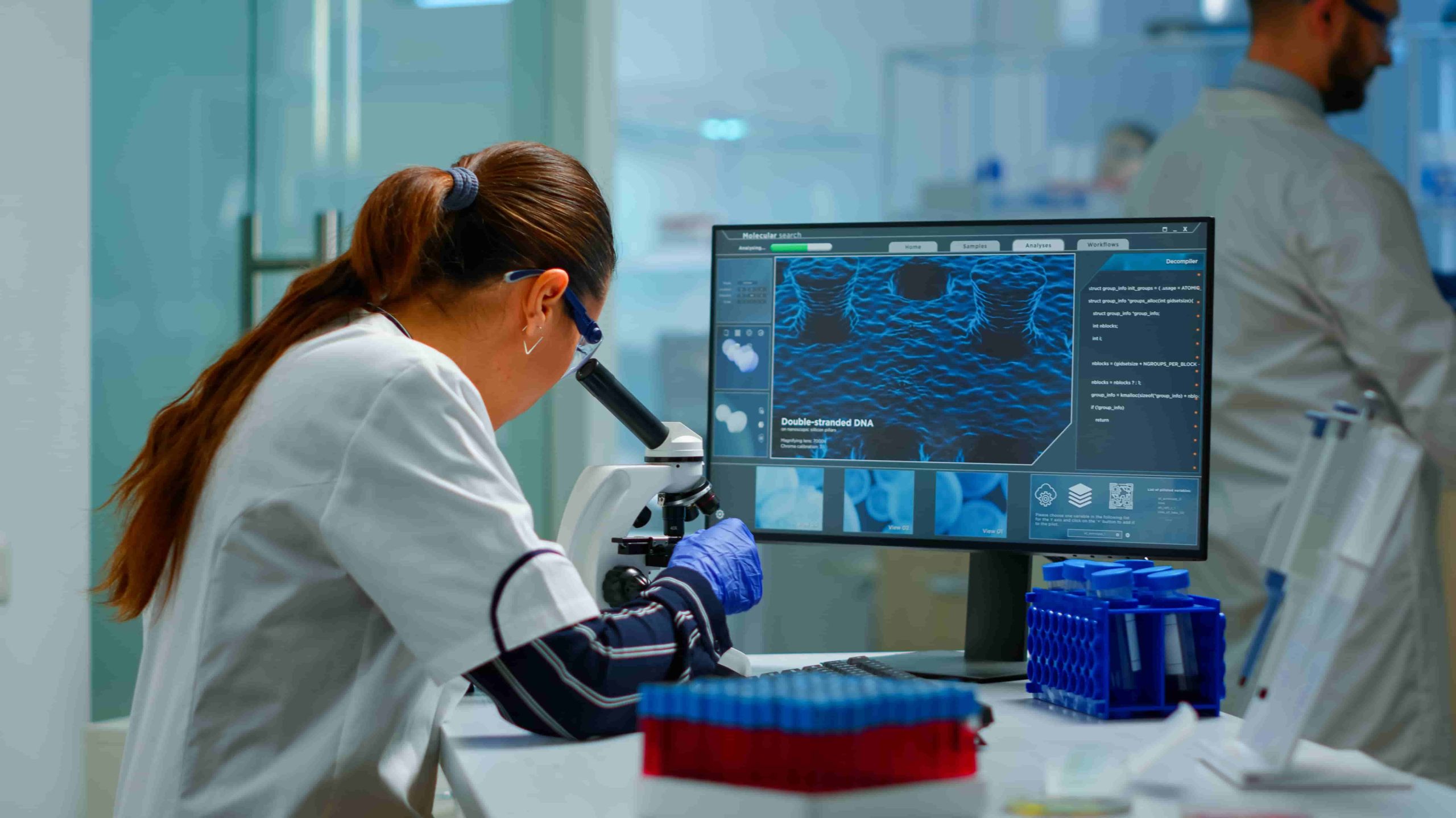
3D Bioprinting Visualization for Personalized Surgical Medicine
3D bioprinting visualization is reshaping personalized medicine by producing patient-specific organ models for precise surgical planning. By integrating advanced imaging with bioprinting technology, surgeons gain unprecedented insights into a patient’s unique anatomy, improving both safety and outcomes. This guide explains how the process works, its benefits, and its future role in healthcare.
What Is 3D Bioprinting Visualization in Medicine?
3D bioprinting visualization combines high-resolution imaging, such as CT or MRI scans, with advanced 3D printing. The result is a lifelike replica of a patient’s organ, allowing surgeons to study, plan, and even rehearse procedures before entering the operating room. This approach blends technology and medicine to deliver care tailored to the individual.
Learn more about Real-World Applications of Quantum Computing in Healthcare.
How 3D Bioprinting Visualization Works
The process of creating a patient-specific model involves three main stages:
-
Medical Imaging – Detailed scans capture the exact structure and condition of the patient’s organ.
-
Digital Modeling – Specialized software transforms these scans into an accurate 3D digital model.
-
3D Printing – Using biocompatible materials, a physical model is printed, ready for surgical preparation.
This streamlined workflow ensures surgeons have a tangible reference for complex procedures, reducing uncertainty.
Benefits of 3D Bioprinting Visualization for Surgeons
Surgeons benefit greatly from 3D bioprinting visualization, as it allows them to:
-
Improve Precision – Models match patient anatomy exactly, supporting accurate surgical planning.
-
Reduce Operating Time – Familiarity with the organ layout speeds up procedures.
-
Enhance Training – Surgical residents and medical students can learn using real-case organ replicas.
Benefits of 3D Bioprinting Visualization for Patients
For patients, 3D bioprinting visualization offers:
-
Fewer Complications – Detailed planning reduces surgical risks.
-
Faster Recovery – Precise procedures mean less trauma and shorter hospital stays.
-
Better Communication – Seeing a model of their organ helps patients understand their condition and surgery.
This personalized approach builds trust between patient and care team, improving the overall healthcare experience.
Applications of 3D Bioprinting Visualization in Surgery
The applications of 3D bioprinting visualization span multiple medical specialties:
Heart Surgery
Cardiac surgeons use printed heart models to plan valve repairs and bypasses. These models reveal structural abnormalities, enabling safer interventions.
Brain Surgery
Neurosurgeons rely on brain models to navigate delicate tissues, avoiding critical regions during tumor removal or aneurysm repair.
Orthopedic Surgery
3D-printed bone models help orthopedic surgeons plan joint replacements and complex fracture reconstructions, ensuring implants fit perfectly.
Challenges in 3D Bioprinting Visualization
While promising, 3D bioprinting visualization faces several hurdles:
-
High Costs – Advanced printers and biocompatible materials remain expensive, though prices are falling.
-
Time-Intensive Process – Printing can take hours or days, which may limit use in emergencies.
-
Regulatory Approval – Models must meet strict safety and accuracy standards before clinical use.
See Simulation of IoT Ecosystems in Real-Time: Key Challenges.
Future of 3D Bioprinting Visualization in Personalized Medicine
The future of 3D bioprinting visualization looks bright, with several advancements on the horizon:
-
Enhanced Materials – New bio-inks and polymers are improving realism and durability.
-
Faster Printing Speeds – Next-generation printers could deliver models within hours.
-
AR/VR Integration – Surgeons could interact with 3D models in virtual environments for enhanced planning.
Conclusion
3D bioprinting visualization is redefining how personalized medicine is practiced, offering safer surgeries, faster recovery, and greater precision. By giving surgeons a hands-on preview of the patient’s anatomy, this technology bridges the gap between planning and practice. As materials improve, costs drop, and speed increases, we can expect 3D bioprinting visualization to become a standard part of surgical care worldwide.
FAQs
What is 3D bioprinting visualization?
It’s the process of creating physical organ models from medical imaging data for surgical planning.
How does it improve surgery?
It provides realistic anatomical models, reducing risks and improving surgical accuracy.
Is it expensive?
Currently yes, but costs are decreasing as technology advances.
What surgeries benefit from it?
Heart, brain, and orthopedic surgeries see the greatest impact.
Author Profile

- Online Media & PR Strategist
- Hello there! I'm Online Media & PR Strategist at NeticSpace | Passionate Journalist, Blogger, and SEO Specialist
Latest entries
 Computer Aided-EngineeringMarch 18, 2026CAE in Sports Equipment Design: Performance & Safety Boost
 AI WorkflowsMarch 18, 2026Nvidia AI Graphics Backlash Explained for Gamers Today
 NetworkingMarch 17, 2026High Speed Networking for Quantum AI Systems Growth
 Vehicle SimulationMarch 11, 2026Physical AI Integration Driving the Future of Smart Cars